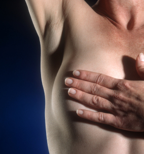
 October is international breast cancer awareness month. Different events and activities are performed during this month in order to raise awareness in women about the gravity of this disease. We would like to educate our patients and raise awareness about the importance of proper and regular examination as to prevent, that is, detect the early stage of this disease.
Breast cancer
Breast cancer is the most common cancer in women, causing every fifth death in women today. Despite the development of science and medicine, this is still a huge health issue. The breast cancer diagnosis is a true “diagnostic jungle”. It is paradoxical that this disease is developed in an organ that is easily accessible to be examined by means of palpation, clinical examinations… Women of all ages are at risk for developing breast cancer. Incidence rate is highest at the age of menopause. Women with positive family history of breast cancer are at higher risk of developing this disease, followed by women with early menarche (first menstruation) and late menopause, women who did not give birth, and women who had their first child after the age of 30. Contralateral breast cancer and endometrial cancer increase the risk of breast cancer development. Genetic (hereditary) factors (gene mutations) also play an important role in breast cancer development.
Regarding Bosnia and Herzegovina, there are between 1000 and 1400 newly diagnosed cases of breast cancer per year, with mortality rate of 500 women!!!
Clinical characteristics of the disease
Main clinical feature of this malignant disease is the presence of a lump in a breast, which develops in about 80% of cases. Depending on which anatomic structures inside a breast are affected, this tumor can cause dimpling or fixation (adherence) of a part of a breast to the front part of the chest wall. Rarely, a nipple discharge can be a sign of breast cancer, and also nipple redness or bloody discharge from a nipple. Breast cancer spreads into the surrounding areas, usually into lymph nodes located in the armpit area.
Breast cancer diagnosis
Palpation test
Palpation is the most important screening method. The breasts are examined while standing, sitting and lying. This type of examination is used to determine size, shape, surface and consistency (hardness), restriction in relation to the surrounding area and change localization. The examination includes palpation of lymph nodes in the armpit area, above and under the collarbone, and comparison with the opposite body side.
Mammography
The main goal of mammography is detection of breast cancer, typically through detection of characteristic masses (e.g. microcalcifications). Mammography is also used for differential diagnosis of different breast processes. Sensitivity and specificity of mammography screening in breast cancer is around 85%, and is increased with the patient’s age. Mammography enables detection of changes smaller than 1cm, and recognition of uncharacteristic indications of breast cancer. Mammography is a contemporary method used for breast cancer screening in women who experience no symptoms (asymptomatic women).
Ultrasound screening
Ultrasound screening is complementary to mammography. Both examinations provide greater accuracy in breast cancer diagnosis. Ultrasound screening is performed in early stages, and it is a method used in targeted breast biopsy cases. Black and white and Color Doppler techniques are used for diagnosis, with multidimensional ultrasound imaging, thus providing better diagnostic accuracy.
Prevention of breast cancer, i.e. reduction of breast cancer development
Breast self-examination
Inspection – looking at your breasts
The first phase of self-examination is visualization (looking at your breasts). Take off your clothes and stand in front of a mirror. Keep your arms close to your body and look at the mirror. Look at your breast and area around them. Pay particular attention to the following:
- Changes to breast size and shape

- Swelling or distortions
- Color change (redness on one or both breasts)
- Nipple position, nipple dimpling
- Nipple color (are both nipples of the same or different color?)
- Breast skin features (is skin smooth or orange-like)
IN FRONT OF A MIRROR: Lift your right arm as if you are reaching for something. Observe if your breast behaves as it did before when raising your arms (position, breast movement, skin tension, etc.). Do the same with the left breast.
Next step: Raise your both arms and keep them behind your head, as if you are about to do sit-ups. Move slowly to the left, i.e. right side and observe whether there are any kinds of changes on your breasts, armpits or area above or under the collarbone.
Keep your arms on your hips and bend forward slightly. You will feel your chest muscle tense. Observe for skin folding, whether it is smooth or not, are there any changes in skin color, etc.
Palpation – exam by touch
For a correct palpation exam you should use your palm, and with the pads of your index finger, middle finger and ring finger make movements over the entire breast. These fingers should be tight, as to avoid skipping a possible lump. Use clockwise circular movements to check your breast. Begin from the outer part towards the nipple. Continue with these movements until you examined the entire breast. The entire time the fingers must be in a firm contact with the breast skin. This is the reason why breasts are never examined “dry”. The best way to do the palpation exam is while showering. If you are not in the shower, rub some oil or body lotion onto your breasts.
It is crucial to examine both breasts and the area surrounding them, including the armpits. Make three circular movements of different intensity on every part: light, medium and firm pressure.
Breast exam should also be done while lying and standing.
Lying position: when lying down on your back put a pillow under either your right or left shoulder, depending on which breast you will examine. Place your arm behind your head. With your other hand examine the breast tissue, as described above.
Standing position: Place your right or left arm (depending on which breast you are about to examine) behind your head, and do the breast exam as described above. In the end, examine your nipples and see if there are any changes to color; then, squeeze the nipple with your fingers and check for discharge.
It is important to perform the exam in both standing and lying positions. Certain changes are more easily detectable while standing, whereas others while lying. By doing so, you will gather more information. In case of any changes, do not panic, but ask for advice and medical help.
NOTE! Self-examination is not a substitute for a clinical examination!!!
Preventive examinations taken in different age and health conditions in women
| Age/health condition | Self-examination | Clinical examination | Ultrasound examination | Mammography |
| 20-35 | 1/month | 1/year | 1/year | No |
| 35-40 | 1/month | 1/year | 1/year | Basic mammography |
| 40-49 | 1/month | 1/year | 1/year | 1 or 2/year |
| 50+ | 1/month | 1/year | 1/year | 1/year |
| Women with genetic risks | 1/month | 1/year | 1/year | 1/year, first mammography in the age of 35 |
| Women after being treated for breast cancer | 1/month | 2/year | 2/year | 1/year |
The Mehmedbašić institute offers service of two, three or four dimensional ultrasound, with black and white and Color Doppler imaging, which, together with the help of professional employees, provides a good-quality clinical breast examination (palpation and ultrasound examination).
The Mehmedbašić genetic laboratory provides services of detecting breast cancer risk by determining gene mutations related to this disease (BRCA1 and BRCA2).


